
In vitro development of a mouse oocyte, from germinal vesicle stage to metaphase II stage. Chromosomes are in red (imaged with fluorescence); spindle, oocyte membrane, and zona pellucida in white (imaged with polarization microscopy)
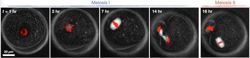
Snapshots of mouse oocyte development. Chromosomes are in red (imaged with fluorescence); spindle, oocyte membrane, and zona pellucida in white (imaged with polarization microscopy)
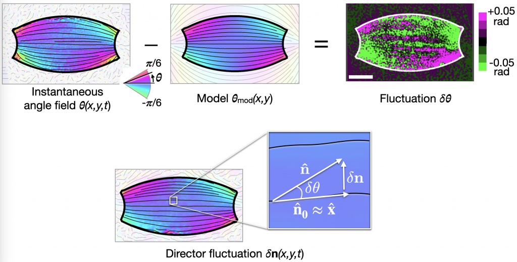
Fluctuations of the microtubule orientation field can be used to probe the mechanics of living oocyte spindles